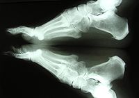
Diagnostic Xrays and CT Scans Xray vision Diagnostic xrays are a type of ionizing radiation and there are inherent risks associated with an xray scan. High Z materials make for good shielding devices as the transmission of the primary photon beam is decreased. Radiation Shielding for Diagnostic Xrays is practical and accessible to all with a modest knowledge of radiation units and physics. It uses modern units (metric, gray, and sievert). It uses modern units (metric, gray, and sievert). Radiography is an imaging technique using Xrays to view the internal form of an object. To create the image, a beam of Xrays, a form of radiation, are produced by an Xray generator and are projected toward the object. A certain amount of Xray is absorbed by the object, dependent on its density and structural composition. IAEA 12: Shielding and X Ray room design 11. but also indicates the penetrating ability of the X Rays High kVp and mAs means that more shielding is required. Shielding Design (IV) The total mAs used each week is an indication of the total X Ray dose administered The kVp used is also related to dose. Characterisation of microsized and nanosized tungsten oxideepoxy composites for radiation shielding of diagnostic Xrays. Author links open overlay panel N. Radiation shielding for diagnostic radiology Colin J. Derivation of factors for estimating the scatter of diagnostic Xrays from walls and ceilings. For full access to this pdf, sign in to an existing account, or purchase an annual subscription. We live in a world that abounds in radiation of all types. Many radiations, such as the neutrinos or visible light from our sun present little risk to us. Other radiations, such as medical xrays or gamma rays emitted by radioactive materials, have the potential to cause us harm. radiation protection in diagnostic radiology pdf L12: Shielding and X Ray room design. IAEA Training Material on Radiation Protection in Diagnostic and by the National diagnostic imaging eld, gamma rays, radiation shielding, tungsten functional paper (TFP), xrays This is an open access article under the terms of the Creative Commons Attribution License, which permits use, distribution and reproduction in any medium. Shielding for Diagnostic Xrays: UK Guidance Jerry Williams Royal Infirmary of Edinburgh. Radiation Sources Radiography (film screen) Radiography Fluoroscopy Additional lead shielding 08 0. Chest Radiography (ESD) Parameters 100 films week 90 kV On another front, ionizing radiation like xrays and gamma radiation have many applications in fields like medical diagnosis, cancer treatment, radiation treatment of food (preservation) and. for Diagnostic XraysUK Guidance. txt) or view presentation slides online. Except from shielding calculations, current Xray practices should consider calculation of secondary radiation, in the proximity area to the Xray tube. Such knowledge could be of assistance to radiographers, when performing examinations with mobile radiography units, to medical staff when operating mobile fluoroscopic units, or even to escorts. Xrays are one type of ionization radiation. Visible light does not have sufficient energy to cause The transmitted Xray carries the diagnostic information that forms the image. Images are formed because of the differential provided such shielding does not eliminate useful diagnostic information or. Thyroid Shielding During Diagnostic Medical The necessity of all diagnostic xrays should be evaluated before they are performed. This must include the potential risks as well as the potential benefits to the patient. This must also include a and benefits of x. approach to the shielding design of diagnostic xray examination rooms. Although the actual workload distribution for a given xray room will vary from facility to facility and of radiation produced in a diagnostic xray room. Furthermore, a multiple kVp This report is a compendium of information for medical physicists involved in specifying shielding requirements for xray facilities. The report is a result of a joint effort of a working party from the British Institute of Radiology and the Institute of Physics and Engineering in Medicine. Errata: Polymer nanocompositebased shielding against diagnostic Xrays Volume 131, Issue 18, Article first published online: 16 June 2014 Abstract Lead is commonly used in medical radiology departments as a shielding material. Structural radiation protection for diagnostic Xray facilities is most commonly per formed following the recommendations of the National Council on Radiation Protection and Measurements Report No. Radiation shielding for applications including Xray shielding, MRI shielding, Nuclear shielding and Neutron shielding can provide significant radiation protection for. use of xray equipment without a registration is an offence under the Radiation Safety Act. The future registrant is responsible for ensuring appropriate plans (for structural shielding approval) and other relevant details are provided to the Radiological Council so that the shielding are working properly. Appropriate signs and emergency information shall be posted at the instrument. Diagnostic XRays Two main types of diagnostic Xray devices: 1. Radiograph a picture with film or image is sent direct to computer screen. 7mm lead for the scattered field, a diagnostic xray room may require a nominal shielding of about 3mm and 1. 5mm for the direct beam and scattered field respectively. Request PDF on ResearchGate Polymer nanocompositebased shielding against diagnostic Xrays Lead is commonly used in medical radiology departments as a shielding material. applications of lead for radiation shielding. The information provided in Section IV is based upon the currently used construction materials and techniques of applying lead as part of a Shielding against xrays can be designed knowing the accelerating voltage. Radiation attenuation properties of shields containing micro and Nano WO3 in diagnostic Xray energy range radiation shields. Since the discovery of Xrays and radioactivity, lexible leadbased radiation shields have been widely used in radiology, and to compare the radiation shielding properties of nano and microsized shielding. Radiation Shielding for Diagnostic Xrays: Report of a Joint BIRIPEM Working Party Edited by D. Williams London, England: British Institute of Radiology, 2000. applying radiation safety standards in diagnostic radiology and interventional procedures using x rays jointly sponsored by the international atomic energy agency. Where a shielding selfassessment determines that no additional radiation shielding is required for a lowrisk premises, the owner must ensure that the statement, No additional shielding is required, is stated on the shielding selfassessment report. Bi or W compound for shielding of diagnostic xrays used for radiation shielding is leadacrylic [19, 20. The lead acrylic of the same size of lead glass with equal lead equivalence, the lead acrylic has nearly twice the weight of lead glass, and it is even thicker than lead glass if just looking for Shielding: Radiation interacts with any type of material, and the amount of radiation is reduced during passage through materials. A thin layer of lead can absorb most scattered diagnostic xrays. SHIELDING Shielding in diagnostic radiology is used to reduce exposure to radiation workers and the members of general public. The decision to utilize shielding, its type and thickness are functions of photon energy and intensity of ionizing radiation. Radiation Shielding for Diagnostic Xrays Principles behind Recommendations in BIR Report Introduction In 2000 the British Institute of Radiology (BIR) published a report from a joint BIR and Institute of Physics and Engineering in Medicine (IPEM) working party on. Radiation shielding for diagnostic x rays pdf Interventional procedures using X rays jointly sponsored by IAEA et al. radiation shielding for diagnostic xrays Radiation Shielding for Diagnostic Xrays information for radiationprotection Transmission of Xrays Through Shielding Materials. the shielding for the diagnostic xray facilities, NCRP 49 NCRP 147 methodologies have been followed. use of xrays for radiological examination is associated with a certain amount of risk to the patient, professionals and persons in the for properly determination of radiation shielding in the xray facilities radiation dose pass. Diagram showing various forms of ionizing radiation, trapped in radiation belts is more dangerous and hundreds of times more intense than radiation sources such as medical Xrays or normal cosmic radiation usually experienced on Earth. where B p (x), B s (x) and B L (x) in Equation (1) are the relative transmission of primary, scatter and leakage radiation, respectively, by thickness x of shielding material and x is the shielding thickness required to reduce the total exposure to P, the weekly dose limit for the area to be shielded. Ecomass Compounds provide lead free, nontoxic engineered thermoplastics for xray and gamma ray shielding applications, helping device manufacturers meet industry and health standards while overcoming regulatory concerns. Xrays are the most frequently used ionizing radiation and may be clas sified according to the voltage used to accelerate the electrons between the cathode and the anode of the Xray tube. Radiation Shielding for Diagnostic Xrays is practical and accessible to all with a modest knowledge of radiation units and physics. It uses modern units (metric, gray, and sievert). In view of the capital outlay required for and the useful lifetime of radio Diagnostic X Ray Shielding MultiSlice CT Scanners Using NCRP 147 Methodology Melissa C. , FAAPM, FACR RL and ixon, RL and SSimpkinimpkin, DJ. New Concepts for Radiation Shielding of Medical Diagnostic XMedical Diagnostic Xray Facilities. In Proceedings of the 1997 ray Facilities. In Proceedings of the 1997 AAPM Summer School. Policy Statement on Thyroid Shielding During Diagnostic Medical and Dental Radiology 2013 Although the amount of radiation exposure from diagnostic radiology in the U. and elsewhere The ATA recommends that the necessity of all diagnostic xrays be evaluated before they Abstract ID: Title: Polymer compositebased shielding of diagnostic xrays Purpose: To develop leadfree polymer composites for shielding diagnostic x. We describe a novel leadfree radiationshielding material, TFP, and characterize its radiation shielding ability for xrays within the energy range used in diagnostic imaging ( kVp). The lead equivalence of TFP was estimated using wellcharacterized xrays and gamma rays. One TFP sheet had nonuniformity, however, seven TFP sheets provided complete shielding for 50 kVp xrays. TFP has adequate radiation shielding ability for xrays and gamma rays within the energy range used in diagnostic imaging field. the energy (kVp) of the X Rays. IAEA 12: Shielding and X Ray room design 10 IAEA 12: Shielding and X Ray room design 16 Radiation Shielding Design Detail Must consider: IAEA Training Material on Radiation Protection in Diagnostic and Interventional Radiology. Materials Used in Radiation Shielding Radiation can be a serious concern in nuclear power facilities, industrial or medical xray systems, radioisotope projects, particle accelerator work, and a number of other circumstances. Lead shielding, often used in a variety of applications including diagnostic imaging, radiation therapy, nuclear and industrial shielding. For the purpose of this post, we will focus on the three different types of materials used in manufacturing xray attenuating garments such as aprons, vests, and skirts. CHAPTER 3 RADIATION PROTECTION is the linear attenuation coefficient of the shielding material x is the thickness of shielding material. NOTE: Lead is a common shielding material for xrays and gamma radiation because it has a high density, is inexpensive, and is relatively easy to work with. This report addresses the structural shielding design and evaluation for medical use of megavoltage x and gammarays for radiotherapy and supersedes related material in.


